According to the World Health Organization (WHO) 2020 report, there were nearly 2.3 million women diagnosed with breast cancer in 2020 alone. Despite significant advances in diagnosis and treatment, nearly 685,000 deaths were recorded globally from breast cancer in 2020 (https://www.who.int/news-room/fact-sheets/detail/breast-cancer). Breast cancer is the top cause of cancer deaths in women around the world and the incidence is rising in every part of the world, including Singapore. WHO has set up a goal of making efforts in reducing the breast cancer mortality by 2.5% annually and saving lives. Many developed countries have shown this to be an achievable goal. The three main pillars are health promotion (breast cancer awareness), early diagnosis, and comprehensive treatment1.
Breast cancer death is directly proportional to the stage of the breast cancer at the time of diagnosis. Patients diagnosed at stage 1 have an overall 5-year survival in more than 90% cases, which steeply reduces to about 20-25% in cases diagnosed with stage 42.
Using low dose x-rays to screen the breasts in mammograms has the ability to detect more than 80% of cancers at an early stage. Doing regular screening mammograms has already proven to reduce breast cancer mortality in many studies performed over decades3.
Common questions and myths about breast cancer
Q. What can I expect when doing a mammogram?
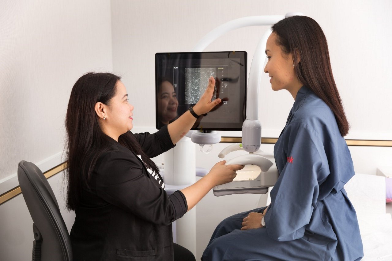
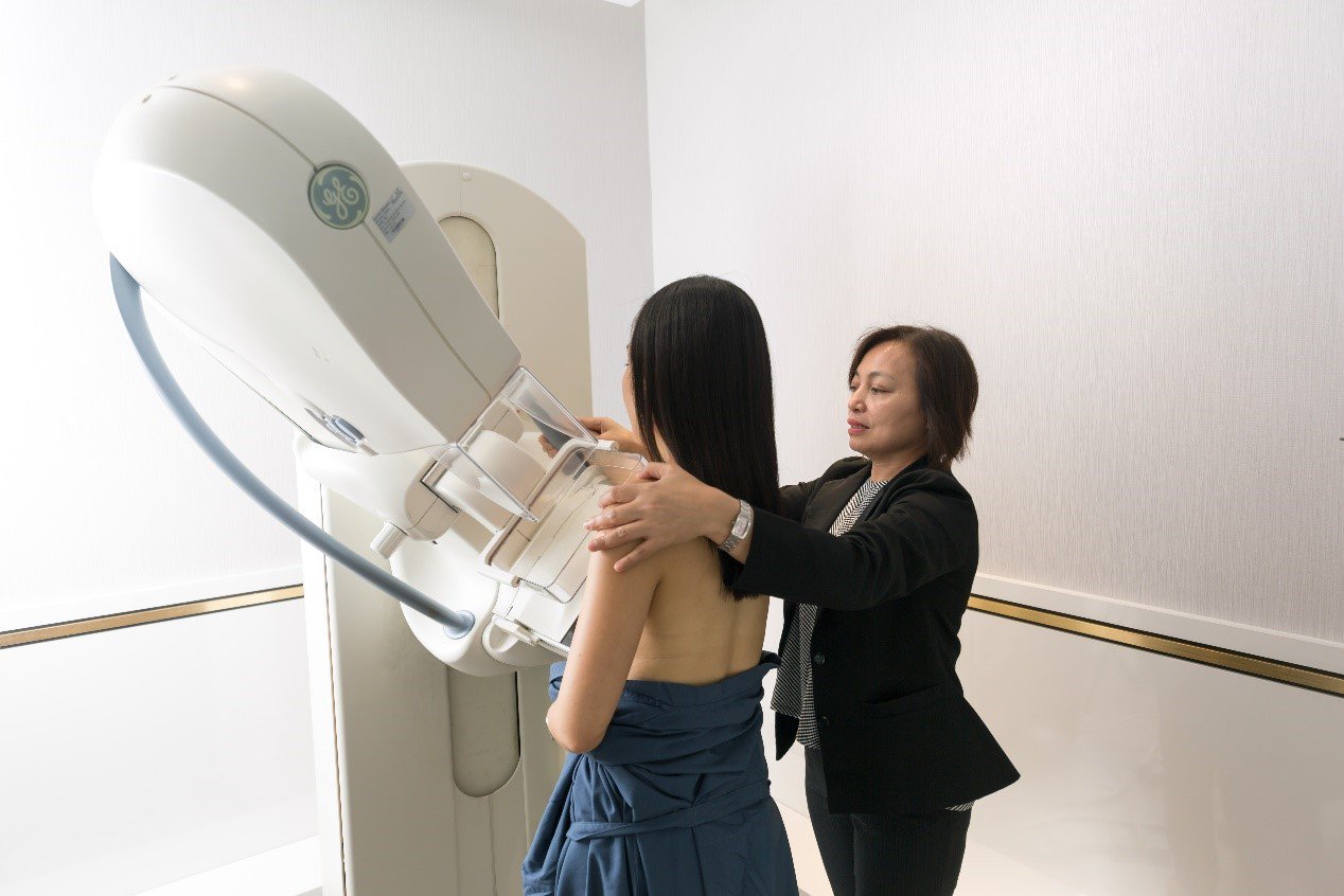
Mammogram is a special kind of x-ray of the breasts. The breast is gently compressed in the machine between the detector plate and compression paddle for less than a minute when the x-ray is obtained. Compression spreads out the breast tissue evenly and increases the chance of cancer detection by reducing the tissue overlap. It also reduces the breast thickness and that in turn reduces the amount of radiation given to the breasts. There might be discomfort during compression; but it is for an extremely short period of time with tangible benefits that overweigh the disadvantage. To reduce the discomfort during compression, it is helpful to schedule the mammogram within the first 15 days of your menstrual cycle for premenopausal females.
To get in-depth details of breasts, two mammograms are performed for each breast as a standard of care. The x-rays (mammographic images) obtained are then read by the specialist who would look for any sign of breast cancer. Comparison with prior mammogram, when available, is of utmost importance as that would allow a slightest change in the appearance to be picked up and thus detect early cancers. When in doubt, the radiologist may request for some additional work up for better clarity and a more comprehensive report to avoid false negative reporting.
Q. Doing a mammogram will cause breast cancer.
With advances in mammography technique and machines, the radiation dose during mammograms is extremely low, in the range of up to 0.4 mSv. It is shown to be well within the safety limits of radiation dose approved by the FDA4. The amount of radiation is so low that the chance of that causing cancer is negligible - estimated to be almost one in a million mammograms. Bear in mind that we are exposed to some sort of background radiation at all times from our surroundings. The radiation from two return international flights from Singapore to USA would mean the same amount of radiation as an annual mammogram. Overall, the advantage of mammograms overweighs the radiation risk and hence one should not avoid mammograms due to fear of radiation.
Q. Mammograms are extremely painful, if a woman who has been treated for breast cancer is nervous about sensitivity and pain during a mammogram, could ultrasound be an alternative?
It is known that some types of breast cancer start with calcifications in the breast and are easily detected on mammograms. On the other hand, it is very difficult to detect breast calcifications on ultrasound. It is not recommended as a standalone screening modality by any of the expert panel around the world5. Ultrasound is best used for breast examination in young women less than 30 years or pregnant or lactating mothers, as mammogram is not preferred at those times. Ultrasound is generally used as an additional modality along with mammogram to increase the chance of cancer detection if the patient has dense breast tissue. Standalone ultrasound is not proven to be the effective modality to detect early breast cancer after the age of 40 years and hence it should not be chosen as an alternative to mammography.
Q. If I do not feel a lump during self-examination, hence I do not have breast cancer.
In more than 85% of cases, breast cancer will produce a palpable lump when they have grown to a significant size6. That means that absence of a palpable breast lump does not rule out cancer in most cases and it will not be wise to go for checking only when one develops a breast lump. That may be a deleterious approach as it may mean diagnosis at a late stage of cancer in many women. The word "screening" itself means that we do the test on 'asymptomatic' women before any signs of cancer are noticed. This would help detect cancer at an early stage and save lives.
Q. Do breast implants have anything to do with increasing breast cancer risk?
Breast implants generally are not found to increase the chance of breast cancer by themselves. Though, it is well known that the presence of breast implants may obscure some parts of breast tissue on mammograms, affecting its sensitivity adversely. Adjuvant use of ultrasound in these cases would add the sensitivity to detect early breast cancer.
Q. I just had a mammogram before and the results were normal, I should be fine and do not need to go for another one.
A negative screening test means that there is no sign of cancer in the study at that time but does not mean that you will not develop cancer in future. Also, in about 15% of women, a small breast cancer may be obscured on mammogram producing false negative results; more so if the breast tissue is dense7. That is why every screening study needs to be performed at regular intervals just like regular health check-ups. By doing so, there is an increased chance of early cancer detection and having improved survival benefits.
According to the Ministry of Health (MOH) recommendations, women between 40 to 50 years of age should be offered mammography annually. After the age of 50 years, performing mammography once every two years is recommended. Various international studies have shown that annual mammographic screening has a better chance of reducing mortality risk compared to biennial mammogram, especially in women with dense breasts8. In Singapore, nearly 70% of women have dense breast parenchyma (tissue) even at post-menopausal age and would benefit from regular mammographic screening.
Q. I do not have a history of cancer in my family and feel healthy, can I wait to have my first mammogram until I am 50?
More than 90% of diagnosed breast cancer patients do not have any family history of breast cancer. Positive family history increases the risk of breast cancer for an individual, but the absence of family history does not guarantee any kind of protection against breast cancer. In the last decade, in Singapore nearly 40% of women diagnosed with breast cancer were below 50 years of age. Incidence of breast cancer at young age is slowly growing and hence it is advisable to start screening annual mammograms at age 40 years and self-breast examination by age 25-30 years9.
Q. My mother had breast cancer when she was in her 40s, when should I start getting a mammogram? Are there any other screening tests I should be getting as well?
Having a first degree relative (mother/father/ sibling) with breast cancer significantly increases the risk of breast cancer; especially if it was diagnosed before 50 years of age of that person. The recommendation is that an annual screening mammogram should be started at least 5 years earlier than the age of the first degree relative at which they were diagnosed3. So, in this case, it would be a good idea to start an annual screening mammogram at the age of 35 years. Also, if the mammogram shows dense breast tissue, then adjuvant ultrasound would increase the chances of cancer detection as the patient is a high-risk individual.
If there are multiple first and second-degree relatives with a history of breast cancer, then it may be wise to see a specialist and check for any genetic predisposition for breast cancer. Calculating lifetime risk and some genetic testing may be done, if advised by the specialist. If proven high risk, then more sensitive screening modality like MRI may be needed in some cases.
Q. Do you recommend breast magnetic resonance imaging (MRI) before surgery for women with breast cancer?
Most studies have shown that preoperative MRI may improve the surgical outcome, but no mortality benefits have been documented in the literature so far. Still, pre-therapeutic MRI has the advantage of better assessment of cancer extent and guiding treatment planning, especially in young women diagnosed with breast cancer or women desiring breast conservation surgery. The need of MRI in a newly diagnosed breast cancer is generally decided by the treating breast surgeon or oncologist depending on each case.
By Dr Niketa Chotai
Consultant Breast Radiologist
MBBS, MD, DNB FRCR, FAMS
Ex-Fellow University of Toronto, Canada
RadLink Diagnostic Imaging Centre
Consultant Breast Radiologist
MBBS, MD, DNB FRCR, FAMS
Ex-Fellow University of Toronto, Canada
RadLink Diagnostic Imaging Centre
This article was contributed by Dr Niketa Chotai, Consultant Breast Radiologist at RadLink Diagnostic Imaging. RadLink is a partner of AIA's Early Detection Screening Benefit. For a list of early detection mammogram screening participating centres, please click here.
References
- Ginsburg O, Yip CH, Brooks A, Cabanes A, Caleffi M, Dunstan Yataco JA, Gyawali B, McCormack V, McLaughlin de Anderson M, Mehrotra R, Mohar A. Breast cancer early detection: A phased approach to implementation. Cancer. 2020 May 15;126:2379-93.
- American Cancer Society. Cancer Facts & Figures 2021. Atlanta, Ga: American Cancer Society; 2021.
- Oeffinger KC, Fontham ET, Etzioni R, et al. Breast cancer screening for women at average risk: 2015 guideline update From the American Cancer Society. JAMA. 2015;314(15):1599-1614.
- Traino AC, Sottocornola C, Barca P, Marini C, Aringhieri G, Caramella D, Fantacci ME. Average absorbed breast dose in mammography: a new possible dose index matching the requirements of the European Directive 2013/59/EURATOM. European radiology experimental. 2017 Dec;1(1):1-8.
- Brem RF, Lenihan MJ, Lieberman J, Torrente J. Screening breast ultrasound: past, present, and future. American Journal of Roentgenology. 2015 Feb;204(2):234-40.
- Koo MM, von Wagner C, Abel GA, McPhail S, Rubin GP, Lyratzopoulos G. Typical and atypical presenting symptoms of breast cancer and their associations with diagnostic intervals: Evidence from a national audit of cancer diagnosis. Cancer epidemiology. 2017 Jun 1;48:140-6.
- Wengert GJ, Helbich TH, Kapetas P, Baltzer PA, Pinker K. Density and tailored breast cancer screening: practice and prediction–an overview. Acta radiologica open. 2018 Sep;7(9):2058460118791212.
- Oeffinger KC, Fontham ET, Etzioni R, Herzig A, Michaelson JS, Shih YC, Walter LC, Church TR, Flowers CR, LaMonte SJ, Wolf AM. Breast cancer screening for women at average risk: 2015 guideline update from the American Cancer Society. Jama. 2015 Oct 20;314(15):1599-614.
- From Singapore Cancer Registry 2018 Annual report: https://www.nrdo.gov.sg/docs/librariesprovider3/default-document-library/scr-annual-report-2018.pdf?sfvrsn=bcf56c25_0